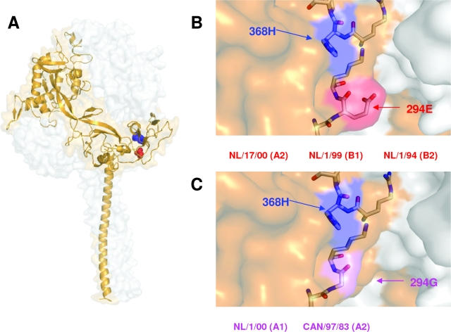
FIG. 4.
Locations of critical residues in a model of HMPV F protein. (A) The model represents the prefusion conformation of the HMPV F trimer, built with the atomic coordinates of the preactive structure of the parainfluenza virus 5 F protein, reported by Yin et al. (19) and using the SWISS-MODEL server facilities (http://swissmodel.expasy.org/). Details of the model-building process will be published elsewhere. One of the monomers is shown as a golden ribbon in which residues 368 (blue) and 294 (red) are shown as balls. (B and C) Partial blowups of the structure showing the lateral side chains of 368H and 294E or 294G. The strains mentioned in the text, which have either 294E or 294G, are indicated below panels B and C, respectively.

