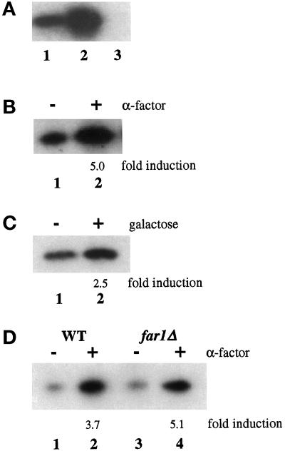
Figure 1.
Phosphorylation and hyperphosphorylation of Clc1p in vivo. (A) Phosphorylation of Clc1p in vivo. Wild-type cells (GPY449, lane 1), wild-type cells overexpressing Clc1p and Chc1p (GPY1118, lane 2), and clc1Δ cells (LSY89.1, lane 3) were grown to midlogarithmic phase and labeled with [32P]Pi at 30°C for 1 h. Clc1p was immunoprecipitated, separated by SDS-PAGE, and analyzed by autoradiography. (B) Clc1p is hyperphosphorylated in the presence of mating pheromone. MATa sst1Δ cells (GPY74-15) were grown at 30°C for 30 min without (lane 1) or with (lane 2) 2.5 μM α-factor. Cells were labeled with [32P]Pi for 30 min, and Clc1p was immunoprecipitated, separated by SDS-PAGE, and transferred to nitrocellulose. After determination of radiolabel incorporation through phosphoimaging, protein content was determined by probing with polyclonal Clc1p antibodies and densitometry. Label incorporated was then normalized to protein content. Values obtained for α-factor–treated cells are divided by values obtained from untreated cells to determine “fold induction” values. (C) Clc1p hyperphosphorylation requires activation of the mating response pathway. Cells (RD680) harboring a galactose-inducible copy of Ste4p were incubated with 2% glucose (lane 1) or 2% galactose (lane 2) for 1 h before incubation with [32P]Pi for 30 min at 30°C. Clc1p was analyzed as described in B. (D) Clc1p hyperphosphorylation is not dependent on cell cycle arrest. Wild-type (WT; FC279, lanes 1 and 2) or far1Δ (FC279, lanes 3 and 4) cells were labeled, and Clc1p was analyzed as described in B.

