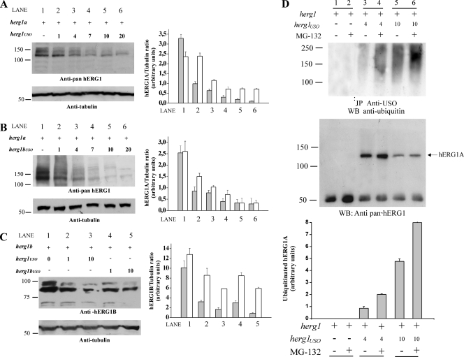
FIG. 5.
Modulation of hERG1A and hERG1B expression by hERG1USO and hERG1BUSO. HEK 293 cells were transiently transfected (+) with herg1a or herg1b alone or with herg1USO and herg1bUSO. Total lysates (5 μg) were run through a 7.5% SDS-polyacrylamide gel, and the following WB was revealed with anti-pan-hERG1 (A and B) or anti-hERG1B antibody (C). Equal loading of proteins in each lane was confirmed by reprobing the membranes with antitubulin antibody (bottom gels). Densitometric analysis was performed as described in Materials and Methods. The positions of molecular mass markers (in kilodaltons) are indicated to the left of the gels. (A) Modulation of hERG1A expression by hERG1USO. Lane 1, cells transfected with herg1a alone; lane 2, cells transfected with herg1a/herg1USO at a 1:1 ratio; lane 3, cells transfected with herg1a/herg1USO at a 1:4 ratio; lane 4, cells transfected with herg1a/herg1USO at a 1:7 ratio; lane 5, cells transfected with herg1a/herg1USO at a 1:10 ratio; lane 6, cells transfected with herg1a/herg1USO at a 1:20 ratio. (Right) Densitometric analysis of mature (gray bars) and immature (white bars) hERG1A protein. (B) Modulation of hERG1A expression by hERG1BUSO. Lane 1, cells transfected with herg1a alone; lane 2, cells transfected with herg1a/herg1bUSO at a 1:1 ratio; lane 3, cells transfected with herg1a/herg1bUSO at a 1:4 ratio; lane 4, cells transfected with herg1a/herg1bUSO at a 1:7 ratio; lane 5, cells transfected with herg1a/herg1USO at a 1:10 ratio; lane 6, cells transfected with herg1a/herg1bUSO at a 1:20 ratio. (Right) Densitometric analysis of mature (gray bars) and immature (white bars) hERG1A protein. (C) Modulation of hERG1B expression by hERG1USO/hERG1BUSO. Lane 1, cells transfected with herg1b alone; lane 2, cells transfected with herg1b/herg1USO at a 1:1 ratio; lane 3, cells transfected with herg1b/herg1USO at a 1:10 ratio; lane 4, cells transfected with herg1b/herg1bUSO at a 1:1 ratio; lane 5, cells transfected with herg1b/herg1bUSO at a 1:10 ratio. (Right) Densitometric analysis of mature (gray bars) and immature (white bars) hERG1 protein. (D) hERG1A ubiquitination after coexpression with hERG1USO and effect of proteasome inhibition. HEK 293 cells were transiently transfected with herg1a alone or with herg1USO at a ratio of 1:4 or 1:10. When needed, transfected cells were exposed to the proteasome inhibitor MG-132 for 60 min (+). Total lysates (1 mg) were immunoprecipitated with anti-USO antibody and run through a 7.5% SDS-polyacrylamide gel, and the following WB was revealed either with an antiubiquitin antibody (top gel) or an anti-pan-hERG1 antibody (bottom gel). The results of densitometric analysis of the ratio between ubiquitinated hERG1A proteins and immunoprecipitated (core-glycosylated) hERG1 are shown in the graph at the bottom of panel D.

