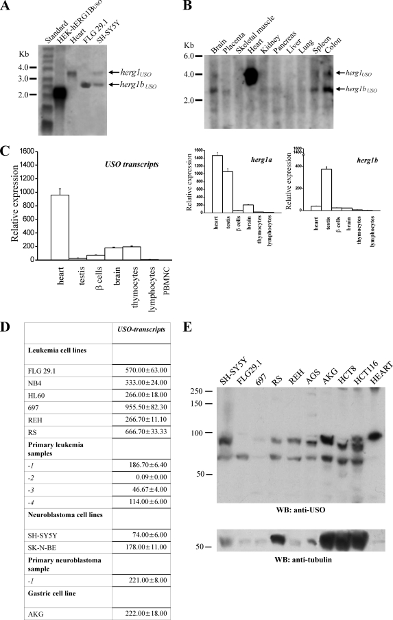
FIG. 6.
herg1USO and herg1bUSO mRNA expression in healthy tissues and cancer cell lines. (A) Northern blot of total RNA extracted from HEK-hERG1BUSO, human heart, FLG 29.1, and SH-SY5Y tumor cell lines, hybridized with a USO exon-specific probe (see Materials and Methods). A single band relative to herg1USO is visible in the heart. The herg1bUSO mRNA in HEK-hERG1BUSO cells is shorter than that in FLG 29.1 and SH-SY5Y cells because the former lacks most of the 5′ untranslated region (see primers used for herg1bUSO cloning, 14). (B) Multiple-tissue Northern blot on poly(A) RNA extracted from different human tissues (Ambion) hybridized with the same USO exon-specific probe. The positions of molecular size standards (in kilobases) are indicated to the left of the gels in panels A and B. (C and D) Quantitative expression of USO-containing transcripts in human healthy cells and tissues (C) and tumor cells and tissues (D) by RQ-PCR. The Sybr green method was used (see Materials and Methods). PBMNC, peripheral blood mononuclear cells. (Right) RQ-PCR of herg1a and herg1b expression on the same RNAs as in panel C. The levels of the various transcripts reported on the y axis are normalized to the level of the corresponding glyceraldehyde-3-phosphate dehydrogenase gene. (D) Quantitative expression of USO-containing transcripts in different human tumor cell lines: acute myeloid leukemias (FLG 29.1, NB4, and HL60), acute lymphoblastic leukemias (RS, 697, and REH), neuroblastomas (SH-SY5Y and SK-N-BE), and gastric cancer (AKG). The quantitative expression of USO transcripts in four different primary leukemias (primary leukemia samples 1 to 4) and in a primary neuroblastoma (primary leukemia sample 1) is also reported. (E) WB of proteins extracted from the tumor cell lines listed above plus a gastric cancer cell line (AGS) and two different colon cancer cell lines (HCT8 and HCT116). The membrane was probed with anti-USO antibody (top gel) and with antitubulin antibody (bottom gel). Note that human heart expressed only the hERG1USO isoform, confirming the Northern blot data. The positions of molecular mass markers (in kilodaltons) are indicated to the left of the gels.

