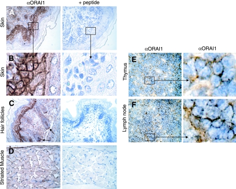
FIG. 1.
Expression of ORAI1 in skin, skeletal muscle, and lymphocytes of wild-type mice. (A to D) Sagittal sections of newborn mice were stained with rabbit polyclonal antibody to ORAI1 followed by peroxidase-labeled secondary antibody and counterstaining with hematoxylin. Where indicated (+ peptide), anti-ORAI1 antibody was preincubated with the immunizing peptide for peptide block. (A, B) ORAI1 staining is present in ectodermally derived epidermal keratinocytes with a pattern indicative of plasma membrane expression (see enlarged area in panel B). (C) ORAI1 is expressed in the mesenchymally derived dermal papillae and dermal sheath of hair follicles. (D) ORAI1 is expressed in the plasma membrane of skeletal muscle. Shown here is a sagittal section of the thoracic diaphragm. (E, F) ORAI1 staining in lymphocytes found in the medulla of thymus (E) and lymph nodes (F) of ∼8-week-old wild-type C57BL/6 mice. α, anti.

