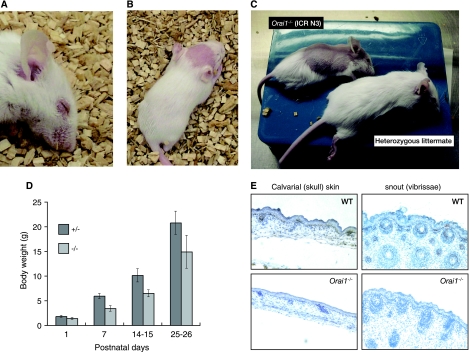
FIG. 3.
Epidermal defects in Orai1−/− mice. (A) Blepharitis (eye irritation) in an Orai1−/− mouse. The blepharitis is even more obvious by 60 days of age, with swelling and reddening of eyelids. (B, C) Hair loss in Orai1−/− mice (15 and 25 days of age, respectively). Hair loss is variable, appearing as completely denuded patches, most frequently on the head. In some cases, the hair loss is progressive, spreading from the head to affect, eventually, a large part of the dorsal skin (C, upper animal) before slowly regrowing to near-normal appearance. (D) Graph of body weights of heterozygous Orai1+/− and homozygous mutant Orai1−/− mice as a function of age. Shown are the averages and standard deviations (error bars) of the weights of 13 Orai1+/− and 8 Orai1−/− mice from a single litter. Results are representative of three litters examined. Orai1−/− mice are consistently 25 to 30% smaller than their heterozygous littermates. (E) Immunohistochemistry of skin from wild-type (WT) and Orai1−/− neonates using ORAI1 antiserum. Tissues were fixed with 4% formaldehyde and embedded in paraffin. Tissue section thickness, 6 μm; ×200, original magnification. Brown (peroxidase) reaction product shows ORAI1 immunoreactivity to be present in keratinocytes in normal skin (upper panels) but absent in Orai1−/− mice. Skin from Orai1−/− mice is thinner, with lower cell density and narrower follicles.

