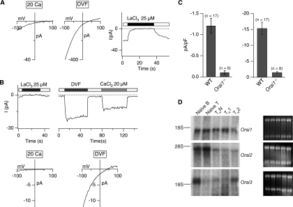
FIG. 6.
Severe impairment of ICRAC in differentiated ORAI1-deficient T cells. (A) Current-voltage relations recorded from differentiated CD4+ T cells derived from wild-type mice in the mixed ICR background in a 20 mM [Ca2+]o and DVF solutions. Store depletion by thapsigargin (1 μM) elicits a Ca2+ current (left panels) and monovalent cation current (center panel) with properties resembling ICRAC in wild-type cells (including inhibition by La3+; right panel). (B) Residual CRAC-like current in differentiated CD4+ T cells derived from Orai1−/− mice. (Top) Currents recorded in a 20 mM [Ca2+]o are blocked by La3+ (left), and residual Na+ CRAC currents are blocked by ∼50% with 20 μM Ca2+ (right), similar to the 50% inhibitory concentration of Ca2+ inhibition in Jurkat T cells. (Bottom) Current-voltage relation in a 20 mM [Ca2+]o and DVF solutions from differentiated CD4+ T cells derived from Orai1−/− mice. Orai1−/− T cells show significant decrease of ICRAC in comparison to the level in wild-type T cells in panel A. In both panels, the bars above the traces indicate the point at which the cell perfusion medium was changed and the duration of time that the indicated substance, stimulus, or [Ca2+]o was present. (C) Summary of peak current densities recorded under the indicated conditions. Error bars represent SEMs. “n” indicates the number of cells analyzed. (D) Results of Northern analysis of Orai transcripts in wild-type naïve B and T cells and differentiated T cells. An amount of 15 μg of total RNA was loaded in each sample. The gels on the right show corresponding 18S and 28S RNA bands as a loading control.

