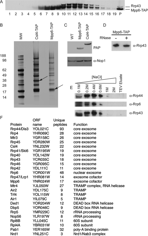
FIG. 3.
Mpp6 physically associates with the exosome. (A) Glycerol gradient analysis of a C-terminal Mpp6-TAP fusion protein. Cell lysate from a strain expressing Mpp6-TAP was loaded on to a 10-to-30% glycerol gradient. Lane 1 is the top of the gradient, and P the pellet. Western blotting was done using peroxidase antiperoxidase, which recognizes the protein A region of the TAP tag, and a polyclonal anti-Rrp43 antibody. (B) Silver-stained gel of affinity purification of Csl4-TAP- and Mpp6-TAP-containing complexes. Arrows indicate the positions of the tagged proteins. MW, molecular weight in thousands. (C) Western analysis of total protein extracts from a wild-type (WT) strain and strains expressing either Mpp6-TAP or Csl4-TAP using peroxidase antiperoxidase (PAP) and an anti-Nop1 antibody. (D) Western blot showing the recovery of the core exosome component Rrp43 with TAP-purified Mpp6 following (+) or without (−) treatment of the IgG-bound complex with RNase A. (E) Western blot showing the elution of the exosome components Rrp43, Rrp44, and Rrp6 from Mpp6-TAP bound to IgG in the presence of increasing salt concentrations. (F) Proteins identified as coprecipitated with Mpp6 by LC-MS. α, anti.

