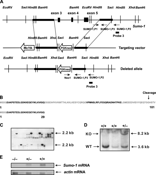
FIG. 1.
Targeted disruption of the Sumo-1 gene. (A) Replacement targeting vector to delete exons 3 to 5 of Sumo-1. The approximate locations of PCR primers used to screen for homologous recombinants and genotypes are shown (arrows) with the original and predicated structures of the gene after homologous recombination. The location of probe 3 used in Southern blotting is also depicted. (B) Amino acid sequence of SUMO-1. Color coding refers to the regions encoded by different exons of Sumo-1. The sequence of the 29-amino-acid long peptide that is potentially encoded by the targeted Sumo-1 gene is shown below. (C) Positive ES clones were found to contain homologous recombination of Sumo-1 by PCR screening using the primers Neo1 and SUMO-1,P2 shown in panel A. (D) Genomic DNA was isolated from two wild-type ES clones (WT) and one representative ES clone with homologous recombination of Sumo-1 (KO), digested with HindIII, and analyzed by Southern blotting. The presence of both 3.2- and 8.6-kb bands indicates the presence of homologous recombination. (E) RNA blot hybridization analysis of samples isolated from testes of wild-type (+/+), heterozygous (+/−), and Sumo-1-deficient (−/−) mice with probes specific to Sumo-1 and α-actin mRNA. The Sumo-1 cRNA probe corresponds to nt 104 to 359 of Sumo-1 mRNA and thus extends from 3′ end of exon 1 until 5′ end of exon 5 of the Sumo-1 gene.

