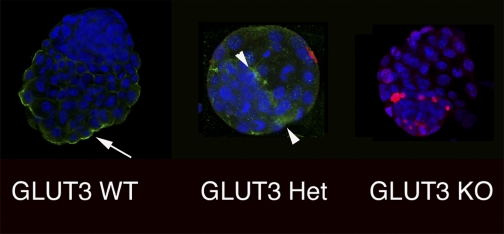
Fig. 4.
Preimplantation embryos demonstrate dual immunohistochemical staining for GLUT3 and TUNEL. A: representative wild type (WT); B: heterozygous (Het); C: homozygous knockout (KO) embryo demonstrating GLUT3 (green) and TUNEL (red) along with nuclear ToPro-3 (blue) staining. WT demonstrates apical distribution (arrow); heterozygous embryo demonstrates punctuate distribution of GLUT3 on apical and basolateral surfaces of the trophectoderm (arrowhead); homozygous embryos demonstrate no GLUT3. TUNEL staining progressively increases from WT to homozygous embryos.

