FIG. 5.
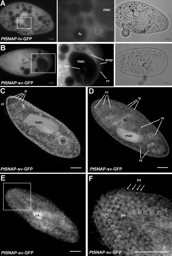
GFP fluorescence in live cells microinjected with a long version (PtSNAP-lv-GFP) (A) and a short version (PtSNAP-sv-GFP) (B), with enlarged details of stained vacuole membranes (middle) and corresponding bright field image (far right). Note that the stained food vacuole (fv) has moved during the objective lens change in the enlargement shown at the right compared to that shown at the left. mac, unstained macronucleus. (B, middle panel) A vacuole (vac) is located on top of the dark appearing macronucleus. The radial canals (rc) and ampullae (amp) of the contractile vacuole system are also weakly stained. (C to F) Confocal image slices (thickness, 1 μm) of fixed PtSNAP-sv-GFP-expressing cells. (C) Median slice showing staining of the membrane of food vacuoles, in the vicinity of trichocysts (tr; the dark, carrot-shaped cortical objects), on cilia (ci) and inside the macronucleus. (D) Median slice showing staining of radial canals and the central contractile vacuole of the contractile vacuole system, between trichocysts and inside the macronucleus. (E) Superficial slice showing staining of dot-like structures and the whole cell surface. cs, cytostome. (F) Enlarged image of a superficial slice showing staining of the whole cell surface and on the regularly arranged parasomal sacs (ps; encircled, between dark trichocysts) but not on trichocyst tips (trt) (indicated by arrows), whose positions can be extrapolated from their regular pattern. Scale bars = 10 μm.

