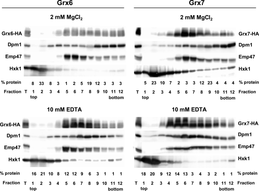
FIG. 1.
Analysis of the association of Grx6 and Grx7 to membrane fractions. Exponentially growing cells (about 3 × 10⁹) in YEPD medium carrying a chromosomally integrated GRX6-3HA (MML897, left) or GRX7-3HA (MML999, right) fusion were employed for obtention of total cell extracts, which were clarified by low-speed centrifugation. The supernatant (fraction T) was subjected to 20% to 60% sucrose gradient centrifugation (5.5-ml total volume of the gradient) in the presence of 2 mM MgCl2 or 10 mM EDTA. Each of the 12 resulting fractions was subjected to Western blot analysis to determine the distribution of Grx6-HA or Grx7-HA, Dpm1 (ER marker), Emp47 (Golgi marker), or Hxk1 (cytosolic marker), by use of adequate antibodies. Ten microliters from each fraction was analyzed in each lane, except for the lane for fraction T, which corresponds to 25 μg of total protein. The relative distribution of total protein along the fractions is also indicated.

