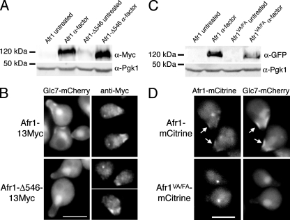
FIG. 4.
Afr1 localization to the necks of projections may be partially dependent on Glc7. (A) Expression of 13Myc-tagged AFR1 confirmed by immunoblot analysis of extracts from AFR1-13Myc (KT2794) and afr1-Δ546-13Myc (KT2795) strains. Protein extracts were prepared from cells growing in logarithmic phase in YPD medium (untreated) or after α-factor treatment. (B) Fluorescence microscopy of Glc7-mCherry and indirect immunofluorescence of Afr1-13Myc in AFR1-13Myc (KT2794) and afr1-Δ546-13Myc (KT2795) strains. (C) Immunoblot analysis of extracts prepared from AFR1-EmCitrine (KT2793) and afr1VA/FA-EmCitrine (KT2792) strains. Protein extracts were prepared from cells growing in logarithmic phase in YPD medium (untreated) or after α-factor treatment. (D) Fluorescence microscopy of Afr1-mCitrine and Glc7-mCherry in AFR1-EmCitrine (KT2793) and afr1VA/FA-EmCitrine (KT2792) strains. Cultures were induced with α-factor. Bars, 5 μm. Arrows mark mating projections where Afr1 and Glc7 colocalize.

