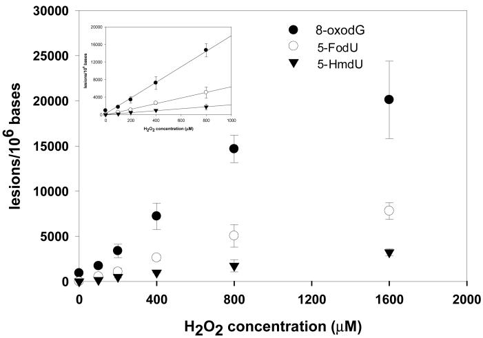
Figure 2.
Cu(II)/ H2O2/ascorbate-induced formation of 8-oxodG (•), 5-FodU (○) and 5-HmdU (θ) in the calf thymus DNA. The values represent the mean ± SD from three independent oxidation and quantification experiments. The inset shows that the yields of the three single-base lesions were proportional to the concentrations of Fenton-type reagents at low dose range.

