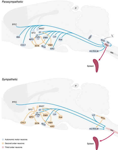
Figure 2. Distribution of pseudo rabies virus (PRV) labeled neurons in different areas of the brain following injection of PRV into the spleen after sympathetic A) or parasympathetic denervation B).
Following different survival times these procedures revealed respectively the first (red) or second order (blue) or third order (brown) neurons that project to the spleen. The upper graph illustrates the areas in the brain that have parasympathetic (pre)autonomic neurons and the lower illustrates those areas that have sympathetic (pre)autonomic neurons.

