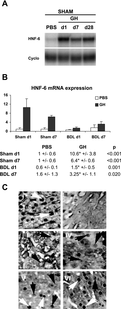
Fig. 1.
Growth hormone (GH) treatment enhances hepatic hepatic nuclear factor (HNF)-6 expression. A: sham-operated mice were treated with PBS or GH for 1, 7, or 28 days (n = 4). Representative RNAse protection assay showed that GH upregulates HNF-6 gene expression. B: mice were treated with PBS or GH for 1 or 7 days following Sham operation (n = 4) or bile duct ligation (BDL; n = 8–10). Real-time PCR analyses showed that HNF-6 was downregulated following BDL whereas GH treatment led to sustained increases in HNF-6 mRNA expression relative to PBS-treated groups. *Significant differences between PBS/BDL and GH/BDL groups with P values ≤0.05. C: representative HNF-6 immunostaining of liver micrographs showed normal level of HNF-6 protein staining, which is localized to hepatocyte nuclei of unoperated liver (I, white arrow showing normal hepatocyte nuclei staining). In contrast, HNF-6 expression was extinguished in hepatocyte nuclei from BDL day 1 (d1) liver as previously reported ( 18) (III, black arrows showing hepatocytes lacking of nuclear staining). Consistent with the recovery in HNF-6 gene expression after 7 days of BDL, faint HNF-6 staining was detected in nuclei of hepatocytes from day 7 (d7) BDL liver (IV, black arrow showing weak staining in hepatocyte nuclei). HNF-6 nuclear staining in BDL days 1 and 7 liver (III and IV) were diminished relative to GH-treated BDL day 1 (V) and day 7 liver; respectively (VI; black and white arrows). C, II: micrographs of Sham-operated liver treated with irrelevant antibody showed no HNF-6 signals.

