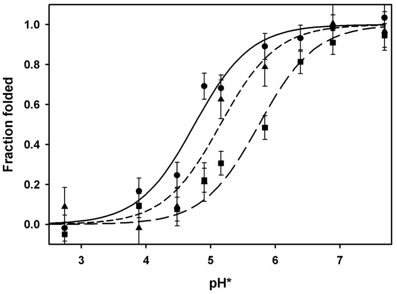
Figure 3. Acid-induced unfolding of bacterio-opsin.
Fraction of folded protein measured by FRET from tryptophan donors to dansyl acceptors. Acceptors: solid line (circles), dansyl groups on Lys 41 (helix B); short dashed line (triangles), dansyl groups on Cys 163 (EF loop); long dashed line (squares), dansyl groups on Cys 222 (helix G). Conditions: 9:1 ethanol:water (v:v), 10 mM ammonium acetate. Titrated with formic and trifluoroacetic acid. pH*: glass electrode measurements uncorrected for junction potentials in 90% ethanol. Lines calculated from pKs in table 1, and error bars indicate standard deviations from lines.

