
Figure 4. Acid-induced unfolding of 5-helix fragments of bacterio-opsin.
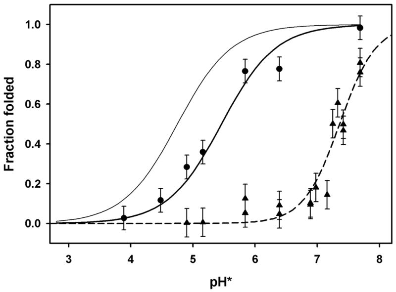
Fraction of folded protein measured by FRET from tryptophan donors to dansyl acceptors. Acceptors: heavy solid line (circles), dansyl groups on Cys 163 (EF loop) of fragment C1 (residues 72-248); dashed line (triangles), dansyl groups on Lys 41 (helix B) of fragment V1 (residues 1-166); thin line, curve fit for Lys 41 label on intact bacterio-opsin (same as solid line in fig. 3). Conditions same as fig. 3.