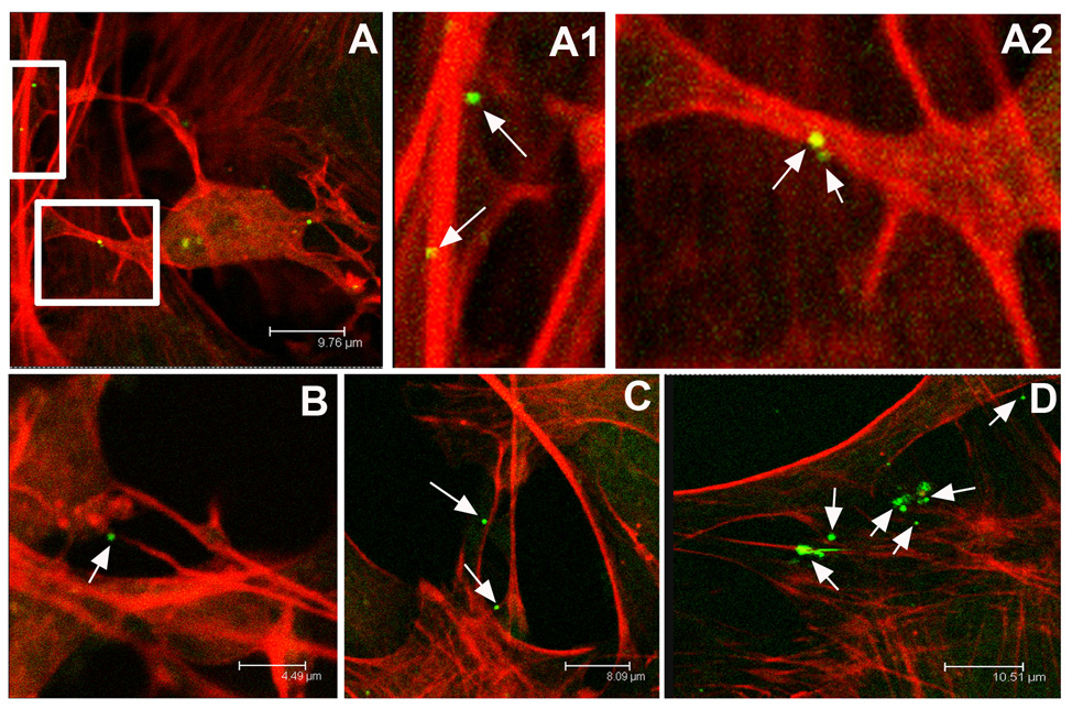
Figure 1.
Confocal microscope images of neurons infected with the neurotropic herpes simplex type 1 virus. Actin networks in P19 cell cultures are visualized in red through cell staining with rhodamine-conjugated phalloidin. Individual viral particles express green fluorescent protein. The virus preferentially associates with long, actin-rich dendritic processes. A, An infected P19 neuron and A1, A higher magnification of a dendrite revealing two green virions attached. A2, A higher magnification of the neuron pictured in A again showing virus attached to red actin-rich dendrites. B, Virus attached to an actin-rich process. C, Multiple HSV-1 virions adjacent to dendritic networks in P19 culture. D, The virus continues to appear bound to actin-rich processes even within complex dendritic networks.

