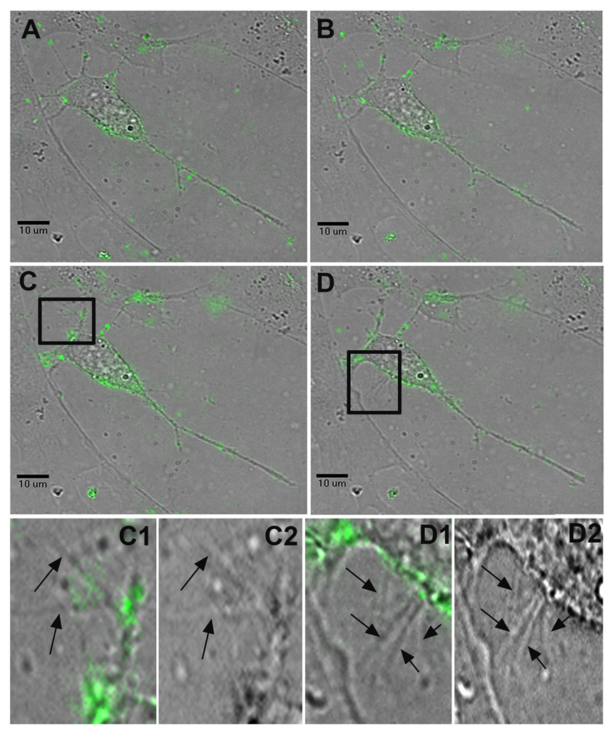
Figure 2.
Surfing of HSV-1 and the induction of dendritic filopodia formation. Still frames taken from a live-cell recording of multiple HSV-1 virions surfing on dendrites towards the soma of a P19 neuron (Supplementary video 1). A, At 5 minutes post-infection, the virus particles have begun to attach to the neuron and its dendrites. B, At 16 minutes post-infection, multiple virions begin surfing along dendrites towards the neuron’s cell body. C, At 25 minutes post-infection, viral particles continue to surf towards the cell body, while significant new dendritic filopodial growth emerges from a previously stable dendrite highlighted in C1, a FITC and brightfield composite image, and C2, a brightfield image alone. D, At 32 minutes post-infection, many viral particles originally found on the neuron’s dendrites have now reached the soma. Another burst of new filopodia appears near the axon hillock and the region highlighted in D1, a FITC and brightfield composite image, and D2, a brightfield image alone.

