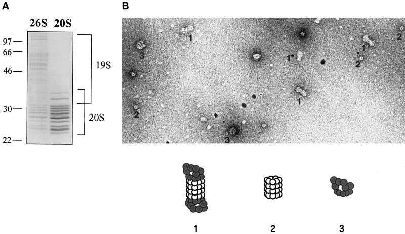
Figure 6.
Characterization of purified yeast 26S proteasomes. (A) Analysis of purified 20S and 26S proteasomes by SDS-PAGE followed by Coomassie Blue staining. Protein size standards (in kilodaltons) are indicated. (B) Electron micrograph of purified 26S proteasome complexes negatively stained with uranyl acetate. Several different species are visible: 26S proteasomes with two PA700 complexes attached at either end (1); 20S proteasome with a single PA700 complex (1*); core 20S proteasomes (2); and complexes that are likely to correspond to free PA700 (3).

