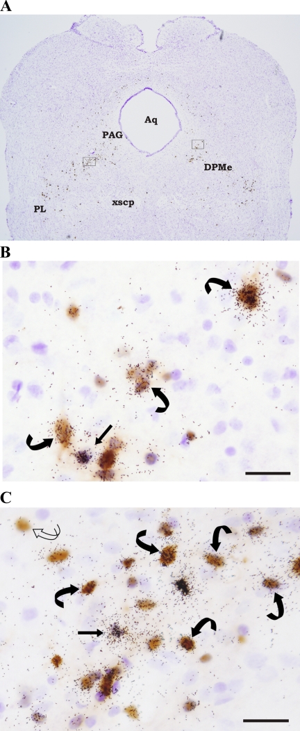
Fig. 3.
A: low magnification photomicrograph of a hamster midbrain section (approximating Plate 69, Ref. 40) double labeled for PRV and MC4-R mRNA. A majority of the midbrain PRV and [MC4-R + PRV] cells were found within the periaqueductal gray (PAG) and deep mesencephalic nucleus (DPMe). Outlined portions of the PAG and DPMe are enlarged in B and C. B: [MC4-R + PRV] cells in the PAG. A single-labeled MC4-R mRNA cell also is shown. C: PRV and MC4-R mRNA labeling in the DPMe. Single-labeled PRV (curved open arrow), single-labeled MC4-R mRNA (straight black arrow), and double-labeled [MC4-R + PRV] (curved black arrows) cells also can be seen in the midbrain DPMe. Arrows indicate representative cells. Aq: aqueduct; PL: paralemniscal nucleus; xscp: decussation of the superior cerebellar peduncle. Bar = 25 μM.

