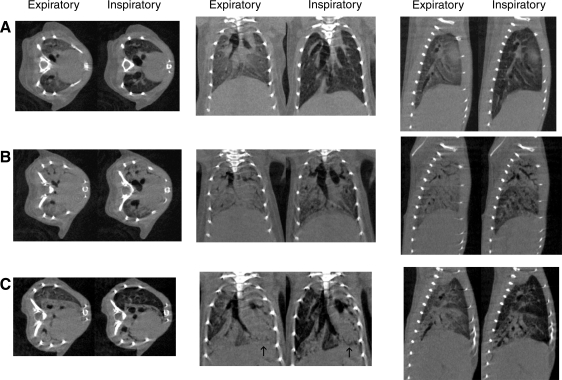
Fig. 3.
Representative micro-computed tomography (micro-CT) images of control mouse (A), week 1 mouse (B), and week 3 mouse (C). Note darkening of lung parenchyma, diaphragmatic depression, and enlargement of the airways present in control mouse (A). In week 1 mouse (B), there is dense consolidation in the expiratory images with ground-glass opacities at end inspiration in the peripheral and lower lung fields with persistent consolidation apically. In addition there is limited movement of the diaphragm, suggesting reduction in lung volumes at end inspiration. Week 3 mouse shows areas of clearing with near normal-appearing lung adjacent to densely consolidated lung that displays minimal change in appearance with lung inflation. Also note asymmetric movement of the diaphragm on coronal images (arrows), suggesting limited inflation in the consolidated left lung.

