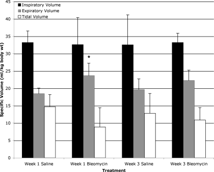
Fig. 5.
Lung parenchymal volumes at end-inspiration, end-expiration, and calculated tidal volumes ± SD for saline- and bleomycin-exposed mice at weeks 1 and 3. Values are normalized for body weight at time of bleomycin or saline administration. End-expiratory lung parenchymal volumes are significantly increased in week 1 bleomycin-exposed mice compared with week 1 saline-exposed mice, *P < 0.042. There were no significant differences between week 3 bleomycin-exposed mice and control mice.

