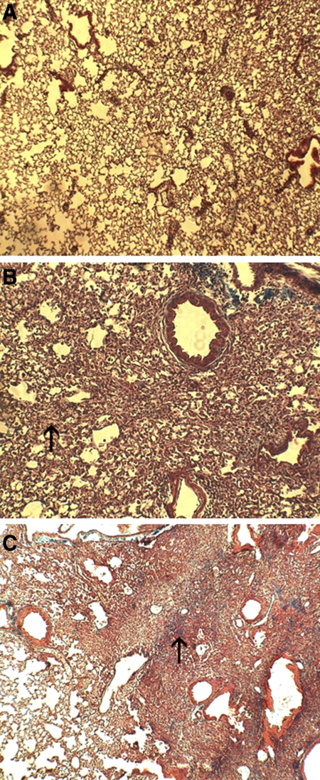
Fig. 7.
Histological samples from control (A), week 1 bleomycin-exposed (B), and week 3 bleomycin-exposed mice (C). Note thickening of the interstitium at week 1 (arrow, B) due to cellular infiltration, with progressive pneumonitis at week 3 characterized by severe architectural distortion and early collagen deposition (arrow, C).

