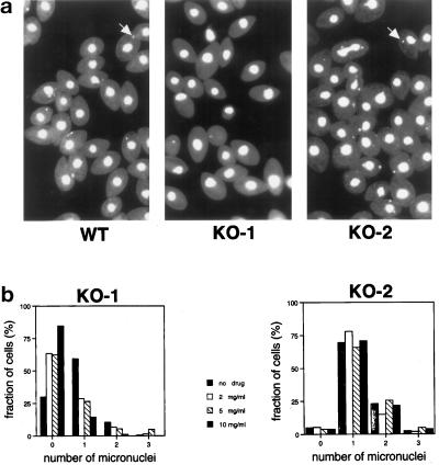
Figure 5.
Many KO-1 cells lack a distinct micronucleus. (a) Fields of cells stained with DAPI and viewed by fluorescence microscopy. Most of the wild-type (B2086) and KO-2 cells contained a micronucleus (examples indicated with arrows), but most of the KO-1 cells did not. The KO-1 and KO-2 cells were grown for several weeks in paromomycin at 10 mg/ml before this experiment. (b) Effects of paromomycin concentration on the number of visible micronuclei per cell in KO-1 and KO-2. Cells were grown in paromomycin at 10 mg/ml and then transferred to medium containing different concentrations of the drug (0–10 mg/ml). After 3 d of vegetative growth, the cells were fixed and stained with DAPI. Approximately 200 cells at each drug concentration were viewed by fluorescence microscopy in a blind protocol. The number of distinct micronuclei in the KO-1 cells was affected by the paromomycin. At high concentrations of the drug, most of the KO-1 cells lacked a visible micronucleus, whereas in the absence of drug, most of the KO-1 cells had one micronucleus. The different paromomycin concentrations did not affect the micronuclear phenotype of the KO-2 cells, demonstrating that the effect in the KO-1 cells was not simply attributable to the drug. The KO-2 cells with two micronuclei included dividing cells in these nonsynchronized cultures.

