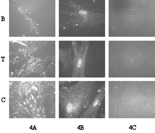
Figure 4.
Immunofluorescent staining for α5 integrin subunit on IMR-90 cells cultured on different Fn-coated substrates for 16 h and cross-linked as in Figure 3. Cells were extracted with either 0.1% SDS (A) or 1% Triton X-100 (B) before staining using an antibody against the α5 cytoplasmic domain. (C) Phase contrast micrographs of Triton X-100–extracted cells. All photographs are at the same magnification (600×) and exposure. More intense staining of the SDS-extracted cells reflects more complete removal of cytoplasmic proteins that can block antibody access to epitopes in the cytoplasmic domain of α5.

