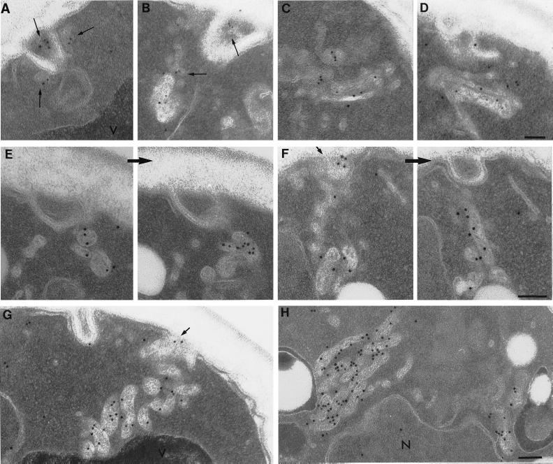
Figure 3.
After exposure to α-factor, Ste2p is located in membrane-bound compartments. Wild-type cells (RH448) were taken after 5 min (A–D), 10 min (E), and 15 min (F–H) of continuous exposure to α-factor. Note small peripheral vesicles (long arrows) and tubular–vesicular compartments to which anti-Ste2p antibodies localize soon after exposure of α-factor (A–D). Note also the perivacuole compartments that localize antibodies directed against Ste2p (F–H). Many of these compartments appear to contain internal membranes and in sequential sections appear as interconnected tubular–vesicular structures (E and F). After 15 min of exposure to α-factor at 37°C, perivacuolar compartments accumulate (G and H). Note in F and G the localization of anti-Ste2p antibodies to tangentially cut invaginations of plasma membrane (F and G, short arrows) as well as to the membrane-bound compartments located proximal to these invaginations. V, vacuole; N, nucleus. Bars, 0.5 μm.

