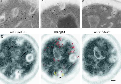
Figure 4.
Ste2p is not located within the cortical actin patch before (A and B) or after (t = 5 min; C and D) exposure to α-factor. (A–C) Examples of double labeling with antibodies against cofilin (15-nm gold particles) and Ste2p (10-nm gold particles) on wild-type cells. Note that Ste2p is not observed in the cortical actin patch (long arrows) but is located in furrow-like invaginations of the plasma membrane (short arrows). (D) Example of adjacent-face double localization using anti-actin (red in merged image) and anti-Ste2p (yellow in merged image) antibodies. The asterisk in D indicates a furrow-like invagination that localizes both anti-Ste2p and anti-actin antibodies; long arrows indicate cortical actin patches (see text). Note that the section in D with anti-Ste2p localization does not appear to be associated with an invagination until the adjacent section is examined. Note also that some furrow-like invaginations are located next to actin patches (A, B, and D). Bars, 0.1 μm.

