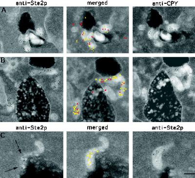
Figure 8.
CPY and internalized Ste2p colocalize in prevacuole compartments. (A and B) Adjacent-face double localization of anti-Ste2p (yellow in merged images) and anti-CPY (red in merged images) antibodies after 15 min of continuous exposure to α-factor at 25°C. Note that the prevacuole compartments appear to be interconnected. (C) Example of adjacent-face double labeling with Ste2p antibodies only. Note that the first section of C shows a grazing cut through a prevacuole compartment that contains vesicles to which Ste2p is localized (arrows). This prevacuole compartment appears to be fusing with the vacuole. V, vacuole. Bar, 0.1 μm.

