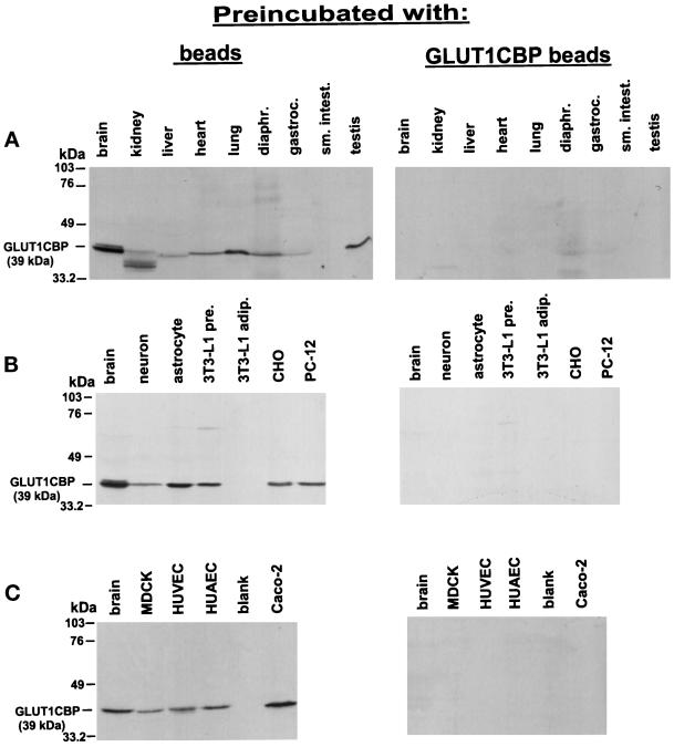
Figure 7.
Analysis of GLUT1CBP protein expression in various tissues and cells. Tissue and cell extracts were prepared as described in MATERIALS AND METHODS. One hundred micrograms of each sample (40 μg for 3T3-L1 preadipocytes and neurons) were separated by SDS-PAGE, and the proteins were transferred to membranes. Analysis of GLUT1CBP expression in tissue samples (A) and in various cell lines (B and C) is indicated with brain controls to allow comparison between blots. Blots were probed with GAb(249–333) antibody raised against the C-terminal 85 amino acids of GLUT1CBP. The antibody was preincubated with Sepharose beads (left) or covalently coupled His6–GLUT1CBP–Sepharose beads (right) before the antibody was added to the membrane. Bound antibody was detected by ECL. adip., adipocyte; diaphr., diaphragm; gastroc., gastrocnemius; pre., preadipocyte; sm. intest., small intestine.

