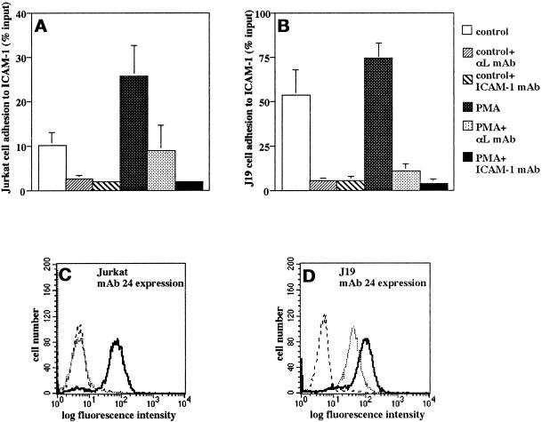
Figure 6.
Differential β2 integrin activation via the α subunit cytoplasmic domains in the mutant Jurkat cell line J19. (A–D) Adhesion to ICAM-1 (A and B) and expression of the mAb 24 epitope (C and D) in J19 cells compared with wild-type Jurkat cells. (A and B) Cells were subjected to adhesion assays on ICAM-1 at 37°C with or without stimulation with PMA (100 ng/ml) for 30 min. For mAb inhibition assays, cells or wells were pretreated with saturating concentrations of mAbs for 30 min on ice. Data are mean ± SD of six independent experiments performed in triplicate. (C and D) Cells were reacted with the CD11α activation epitope reporter mAb 24 in the presence of 1 mM Ca2+ and 1 mM Mg2+ (dotted line) or 5 mM Mg2+ and 2 mM EGTA (bold line) and analyzed by flow cytometry. Staining with an isotype control mAb is shown (stippled line). Shown is a representative experiment.

