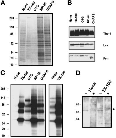
Figure 2.
Kinase activities in GPI-enriched membranes isolated using different detergents. Buoyant, GPI-enriched membranes (0.5 ml, equivalent to ∼25 × 106 cells) from the same cell homogenate, untreated (None) or after treatment with indicated detergents (1% TX-100, 60 mM OTG, 0.5% NP-40, or 20 mM CHAPS), were diluted in TKM buffer, and the GPI-enriched membranes sedimented at 250,000 × g for 4 h were suspended in kinase buffer. (A) Total protein profile after silver staining. (B) Western blot detection of Thy-1, Lck, and Fyn in 20 μl of the GPI-enriched membranes shown in A. (C) Phosphoprotein profile after in vitro phosphorylation in the presence of [γ-32P]ATP as described in MATERIALS AND METHODS. Phosphorylated proteins were separated in 5–20% SDS-PAGE gradient gels and detected by autoradiography after 4.5 h of exposure. For comparison a 20-h exposure of the first two lanes (none and TX-100) is shown in the right panel. Data representative of three independent experiments are shown. (D) Western blot detection of tyrosine-phosphorylated proteins with 4G10 anti-phosphotyrosine mAb after in vitro kinase assay on untreated and TX-100–treated GPI-enriched membranes in the absence (−) or presence (+) of 50 μM cold ATP. The hyperphosphorylated kinase bands are marked by arrowheads.

