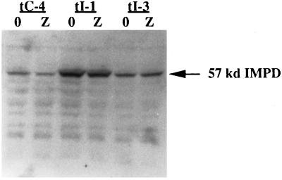
Figure 3.
Increased IMPD protein levels in p53-inducible, impd transfectants. Immunoblot analysis of IMPD protein in soluble extracts prepared from p53-inducible, vector transfectant (tC-4), and impd transfectant (tI-1, tI-3) cells after 2 d of growth under either inducing (Z, 75 μM ZnCl2) or noninducing conditions (0, 0 μM ZnCl2). Forty micrograms of total protein from each extract sample was analyzed as described in MATERIALS AND METHODS with an anti-IMPD antiserum that recognizes both mouse and hamster IMPD proteins. Arrow, the migration position of mouse and hamster IMPD protein.

