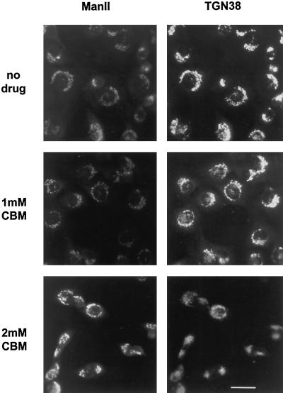
Figure 10.
Distribution of mannosidase II and TGN38 in CBM-treated cells. NRK cells were prepared for immunofluorescence microscopy as described in Figure 4 except that the cells were double-labeled with antibodies to mannosidase II (left) and TGN38 (right).

