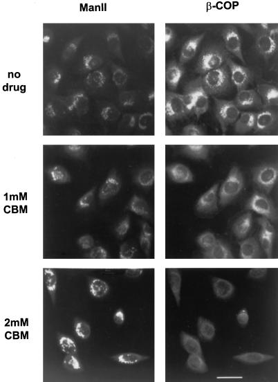
Figure 4.
Distribution of mannosidase II and β-COP in CBM-treated cells. NRK cells grown on glass coverslips were incubated with the indicated concentration of CBM at 37°C for 90 min. The cells were then fixed and double-labeled with antibodies to mannosidase II (ManII; left) and β-COP (right) followed by FITC- and TRITC-coupled secondary antibodies as described in MATERIAL AND METHODS. Bar, 10 μm.

