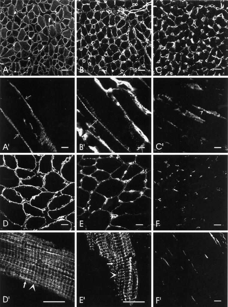
Figure 7.
Spectrins in P7 and adult rat skeletal muscle. Cross-sections (A–F) and longitudinal sections (A′–F′) of 7-d-old (A–C, A′–C′) and adult (D–F, D′–F′) rat diaphragms were labeled with polyclonal antibodies to β-spectrin (A, A′, D, and D′), α-fodrin (B, B′, E, and E′), and β-fodrin (C, C′, F, and F′). The labeling of β-fodrin seen in C and C′ is mostly in capillaries, which are also labeled by anti-α-fodrin (B and B′), but not by anti-β-spectrin. Labeling of the sarcolemma by anti-β-fodrin at P7 is barely detectable. However, some residual punctate labeling could still be seen in longitudinal sections (e.g., arrowheads in C′). By contrast, β-spectrin and α-fodrin are readily apparent at the sarcolemma of P7 muscle, where they are present at punctate structures typical of costameres (e.g., arrows in A′ and B′). β-Spectrin is seen over both the Z lines and M lines in adult muscle (D′, Z line indicated by arrow; M line indicated by arrowhead), with the labeling pattern split into two lines over the Z line. α-Fodrin also localizes over the Z line, but does not show a split there; it is present at M lines to a much lesser extent (E′). Note also that labeling by anti-β-spectrin but not by anti-α-fodrin is present in longitudinally oriented structures, as previously reported (Porter et al., 1997). Bars: A–F, D′–F′, 20 μm; A′–C′, 10 μm.

