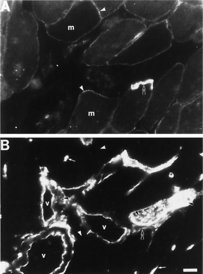
Figure 8.
β-Fodrin and β-spectrin in adult rat skeletal muscle. Cross-sections of adult rat diaphragm were double-labeled with monoclonal antibodies to β-spectrin (A) and polyclonal antibodies to β-fodrin (B). Although anti-β-fodrin antibodies label the blood vessels (v) and nerves (n) brightly, β-fodrin was not detected at the sarcolemma of skeletal myofibers (white arrowheads). β-Spectrin was found at the sarcolemma (A, white arrowheads) and was particularly concentrated at the neuromuscular junction (B, double black arrowhead). Capillaries are indicated with small white arrows; β-fodrin at the membrane of a cell in the blood vessel wall is indicated with a small black arrow. Nerve fibers are indicated with single black arrowheads. m, muscle fiber; v, blood vessel. Bar, 20 μm.

