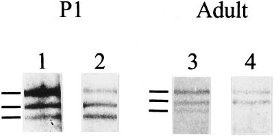
Figure 9.
Immunoprecipitation of spectrin and fodrin from skeletal muscle using anti-fodrin antibodies. Spectrin and fodrin were immunoprecipitated from homogenates of P1 (lanes 1 and 2) and adult skeletal muscle (lanes 3 and 4) using anti-α-fodrin (lanes 1 and 3) or anti-β-fodrin (lanes 2 and 4) antibodies. The immunoprecipitated products were electrophoresed on 5% acrylamide gels and visualized by silver staining. The immunoprecipitated proteins are marked by lines to the left of each set. From top to bottom, they are: α-fodrin, β-fodrin, and muscle β-spectrin.

