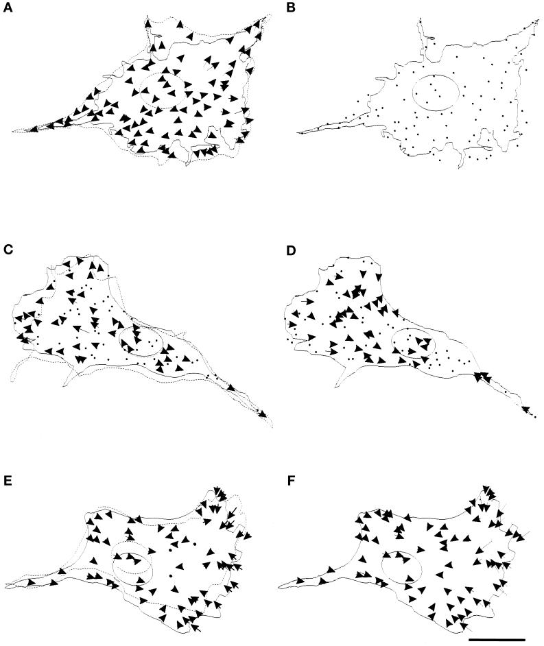
Figure 4.
Effects of drugs on mechanical forces generated by the cell. (A) A cell and beads in the underlying substrate were observed in the presence of 2 μM cytochalasin D for 30 min. The cell stopped forward movement and retracted its boundary (from dotted to solid line), while all the beads moved away from the center of the cell, suggesting the relaxation of forces throughout the cell. (B) The cell was then detached by trypsin to detect any residual deformation at steady state. No bead movement was detected. The dots represent the stationary positions of the beads. (C–F) Similar experiments were performed with 20 μM KT5926 (C and D) and 1 μM nocodazole (E and F). A fraction of thebeads moved away from the center of the cell upon treatment with KT5926, as shown by arrows (C), while others maintained their positions, as shown by dots. No newly developed inward force was detected. The rate of forward movement of the nucleus was reduced by 75% (from dotted to solid line), while the protrusion of the leading edge appeared similar to that in control cells. The forces maintained a radial pattern as in control cells but with an average magnitude reduced by 80–90% (D). Nocodazole had no apparent effect on the overall pattern of forces as compared with that in control cells (E and F). Bar, 20 μm.

