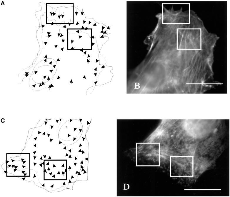
Figure 5.
Distribution of actin filaments and myosin II in relation to strong substrate deformations in the anterior region. (A and C) Cells and beads in the underlying substrate were observed for 20 min. Movements of the beads are indicated by arrows. Dotted and solid lines indicate the starting and ending positions, respectively, of the cell boundary and nucleus. (B and D) The cells were then fixed and stained with rhodamine–phalloidin (B) or antibodies against myosin II (D) to determine the relationship between actin–myosin II organization and mechanical forces. Actin filaments are distributed similarly in areas of large and small substrate deformations (A and B, rectangles). Fine actin bundles are present throughout most regions of the cell. In addition, the concentration of myosin II was similar in regions of strong and weak forces (C and D, rectangles). Bars, 20 μm.

