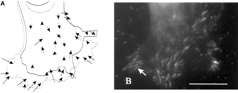
Figure 6.
Distribution of vinculin in relation to large substrate deformations in the anterior region. (A) A cell and beads in the underlying substrate were observed for 12 min. Movements of the beads are indicated by arrows. Solid and dotted lines indicate the starting and ending positions, respectively, of the cell boundary and nucleus. (B) The cell was then fixed and immunofluorescence stained for vinculin to determine the relationship between vinculin organization and mechanical forces. Vinculin is concentrated at elongated plaque structures in the region of strong mechanical forces (B; arrow). Bar, 20 μm.

