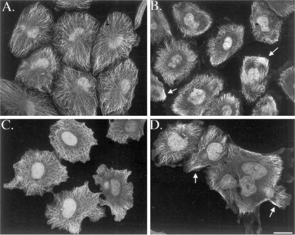
Figure 4.
Arrangement of microtubules in A-498 kidney cells (A, B) and Caov-3 ovary cells (C, D) in interphase in the absence (A, C) or presence (B, 100 nM; D, 30 nM) of taxol (4-h incubation). Cells were stained with an antibody to α-tubulin and imaged using confocal microscopy. A single optical section is shown. Microtubules retracted slightly from the plasma membrane and formed occasional bundles (arrows in B and D) after taxol incubation. Bar, 20 μm.

