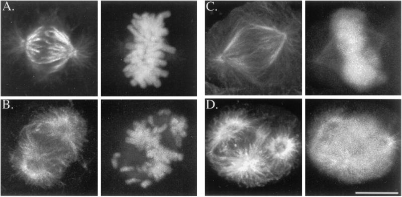
Figure 5.
Microtubules in mitotic A-498 kidney cells (A, B) and Caov-3 ovary cells (C, D) in the absence (A, C) or presence (B, D) of taxol (100 nM and 30 nM, respectively). Cells were incubated with taxol and stained for microtubules (left panels) as described for Figure 4. They were subsequently stained with propidium iodide for DNA (right panels of each pair), and an extended focus series using confocal microscopy was collected. (A, B) Control spindles are bipolar, with congressed chromosomes forming a compact metaphase plate midway between the poles. After 4-h incubation with taxol, spindle morphology is dramatically altered. Many spindles are multipolar (D) with uncongressed chromosomes (B and D). Bar, 10 μm.

