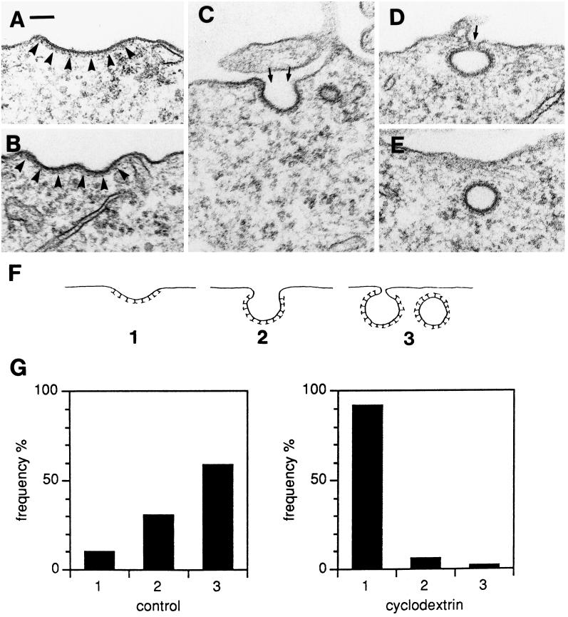
Figure 10.
Examples of clathrin-coated pits at various degrees of invagination (A–D); E shows an apparently free, clathrin-coated vesicular profile that, however, may be surface-connected in another plane of sectioning. The clathrin coat is indicated by arrowheads in A and B. The small arrows in C and D indicate the necks connecting the pits to the exterior. Bar, 100 nm. (F) For quantification, the coated pits and apparently free vesicles (A–E) were subdivided into three types. (G) Coated pits in control HEp-2 cells and cells treated with 10 mM MβCD for 15 min (approximately 200 coated pits in each experiment) were scored at random and classified as 1, 2, or 3 as defined in F. The relative frequency of the different types of coated pits is shown. Bar, 100 nm.

