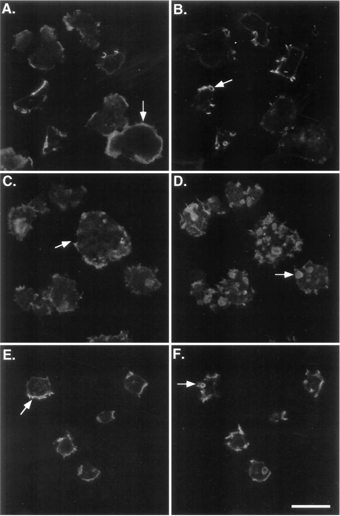
Figure 5.
Expression of full-length myoB rescues the myoA−/B− F-actin defect. Shown are confocal images of rhodamine-phalloidin-stained cells that have been allowed to attach to a substrate for 15 min. Images of the bottom (A, C, and E) and top (B, D, and F) of the cells are presented. The pattern of F-actin distribution in the Ax3 (A and B), myoA−/B− (C and D), and double mutant myoB-expressing strains (E and F) are shown. Examples of the peripheral band staining at the base of the cells (A and E) or brightly staining peripheral spots of F-actin on mutant cells (C) and apical crowns (B, D, and F) are indicated with arrows. Bar, 10 μm.

