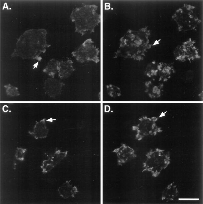
Figure 6.
The F-actin distribution remains abnormal in the myoA−/B− cells expressing mutant forms of myoB. The distribution of F-actin, as determined by rhodamine-phalloidin staining of cells attached to a substrate for 15 min, in the myoA−/B− cells expressing either myoB/SH3− (A and B) or myoB-S332A (C and D) is shown. Confocal images were taken at the bottom (A and C) and top (B and D) of the cells. Examples of the peripheral band staining at the base of the cells (A) or brightly staining peripheral spots of F-actin in mutant cells (A and C) and apical crowns (B and D) are indicated with arrows. Bar, 10 μm.

