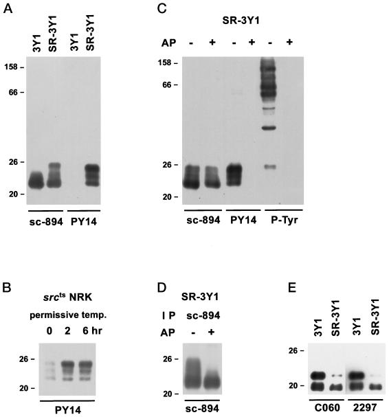
Figure 1.
Western blots using anti–caveolin-1 antibodies and/or PY14. (A) Comparison of 3Y1 and SR-3Y1. Anti–α-caveolin-1 antibody (sc-894) reacted with both samples, but PY14 labeled the latter sample alone. The reaction for SR-3Y1 was broad but with prominent bands at 22, 23–24, and 25 kDa. (B) srcts NRK cells. Reaction with PY14 became detectable only after the cell had been transferred to the permissive temperature. (C) Effect of alkaline phosphatase treatment of nitrocellulose blots of SR-3Y1 lysate. After dephosphorylation, the reactivity with PY14 and anti-phosphotyrosine was lost, whereas the reaction with sc-894 remained. (D) Effect of alkaline phosphatase treatment on immunoprecipitates from SR-3Y1 lysate. Immunoprecipitates obtained with sc-894 were dephosphorylated before being subjected to SDS-PAGE and reacted with sc-894 on blots. Bands above 22 kDa were lost after dephosphorylation. (E) Reactivity of monoclonal anti–caveolin-1 antibodies (clones C060 and 2297) with 3Y1 and SR-3Y1. The antibodies recognize both α and β isoforms in 3Y1 samples, but in SR-3Y1 ones the reaction with the α isoform is much weaker than that with the β isoform.

