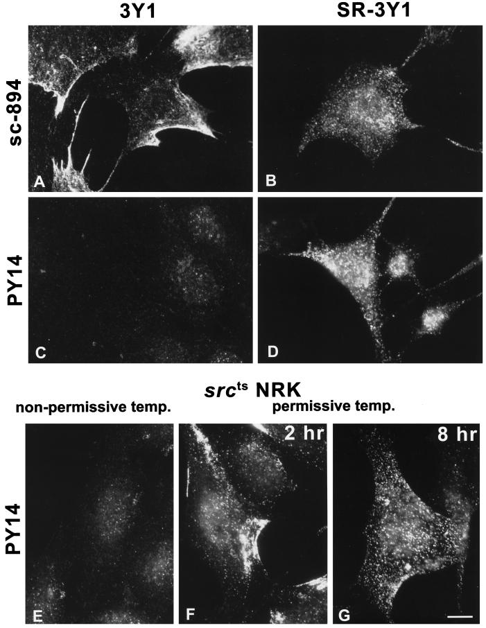
Figure 2.
Immunofluorescence microscopy. (A and B) sc-894 intensely labeled both 3Y1 (A) and SR-3Y1 (B) cells. Note that the labeling occurs as peripheral patches in 3Y1 but as randomly distributed dots in SR-3Y1 cells. (C and D) PY14 does not bind to 3Y1 (C) but labels SR-3Y1 cells in a similar pattern as sc-894 (D); note that some dots are apparently larger than others. (E–G) Most srcts NRK cells were negative with PY14 at 39°C (nonpermissive temperature) (E), but after the temperature was lowered to 33°C (permissive temperature), they showed intense labeling in peripheral patches at 2 h (F) and randomly distributed large dots at 8 h (G). Bar, 10 μm.

