Abstract
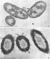
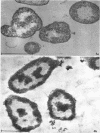
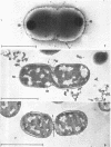
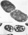
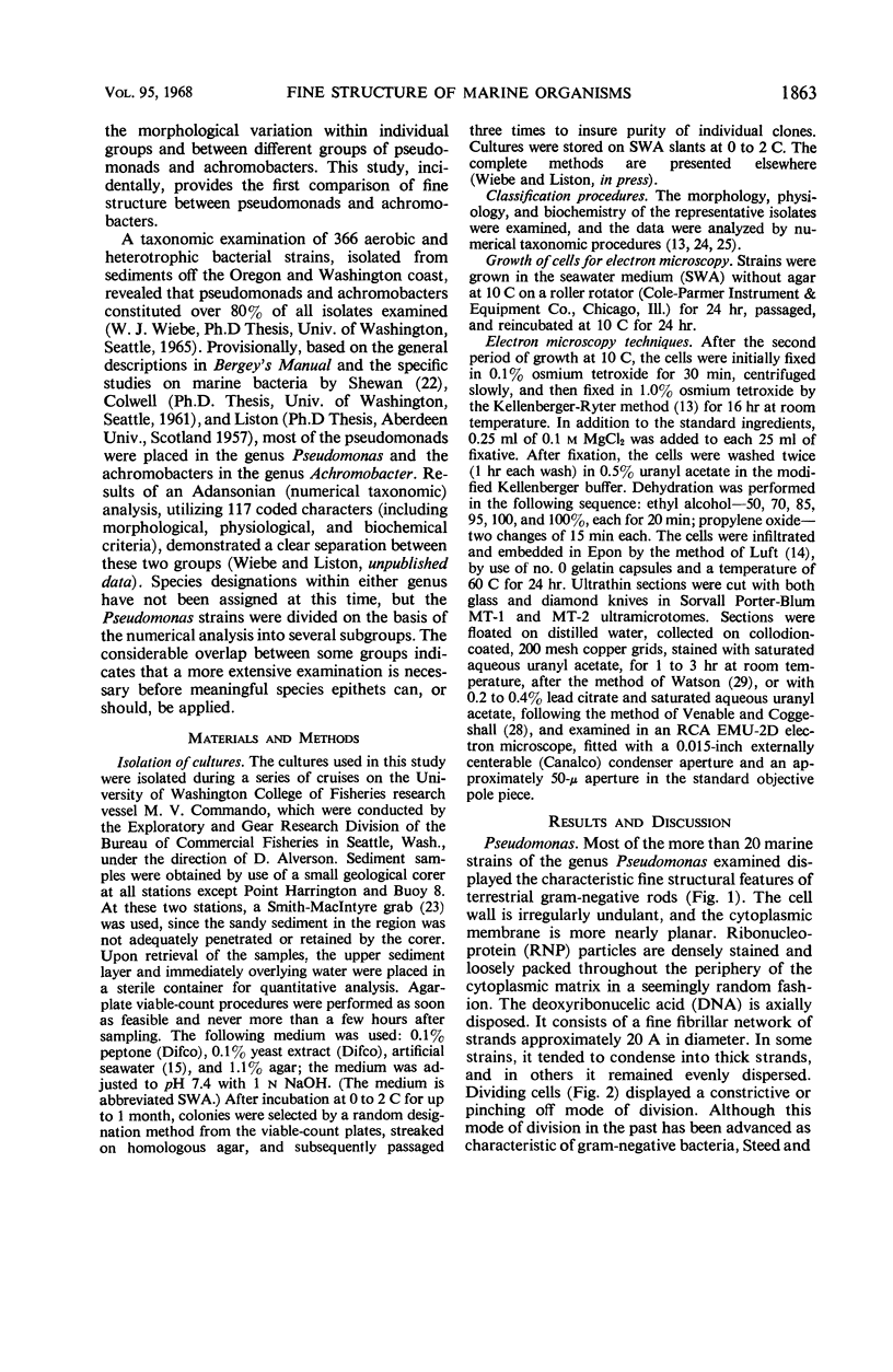
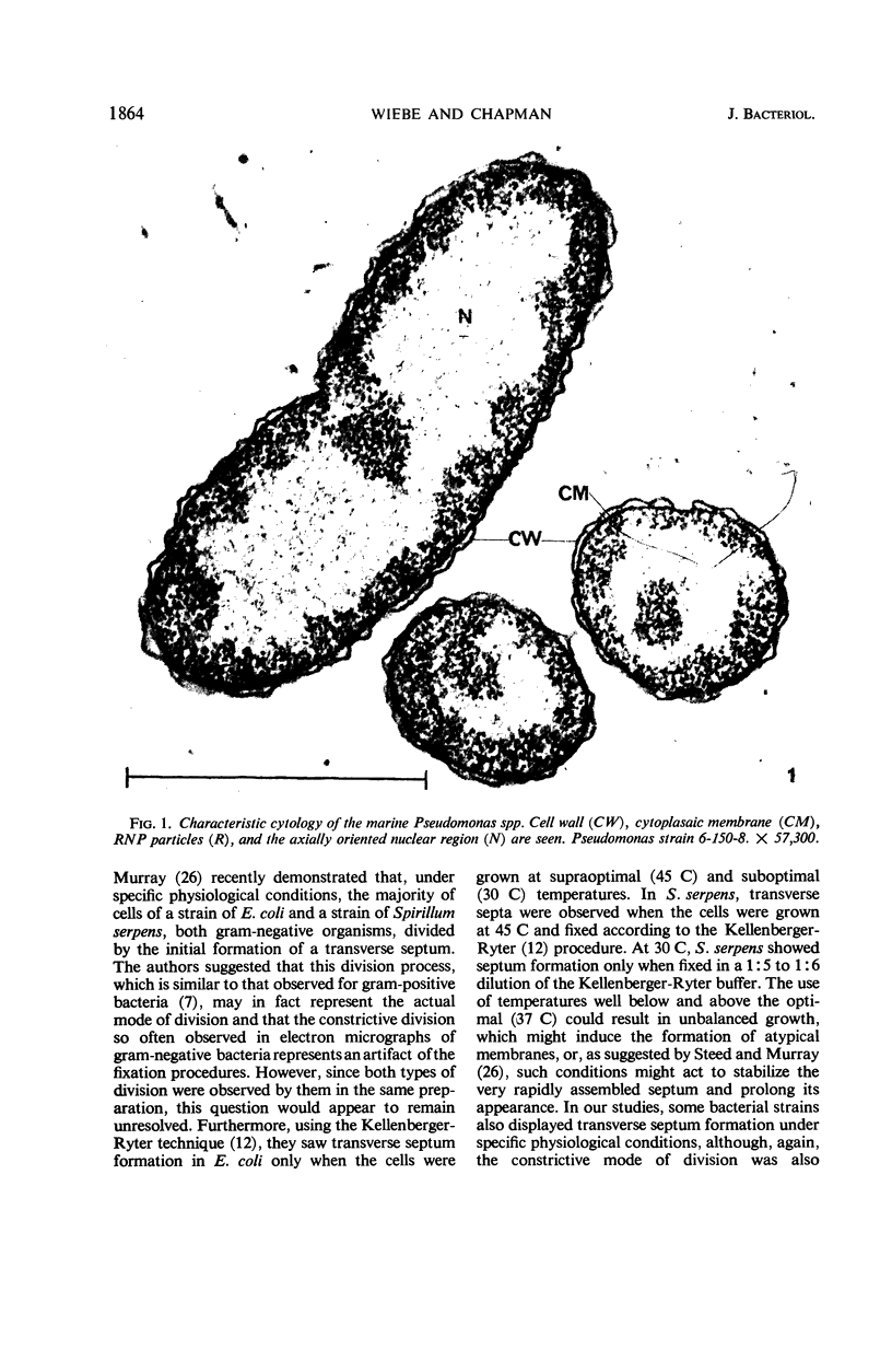
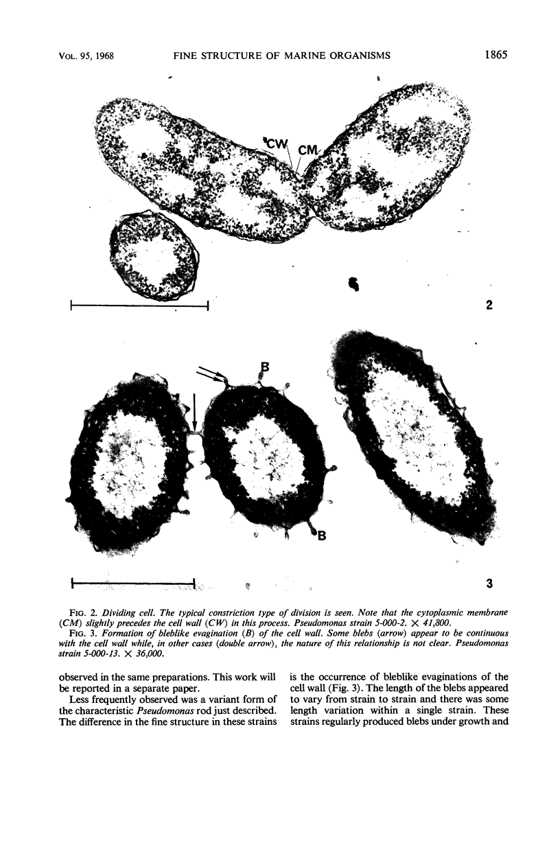
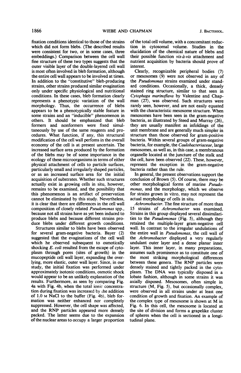
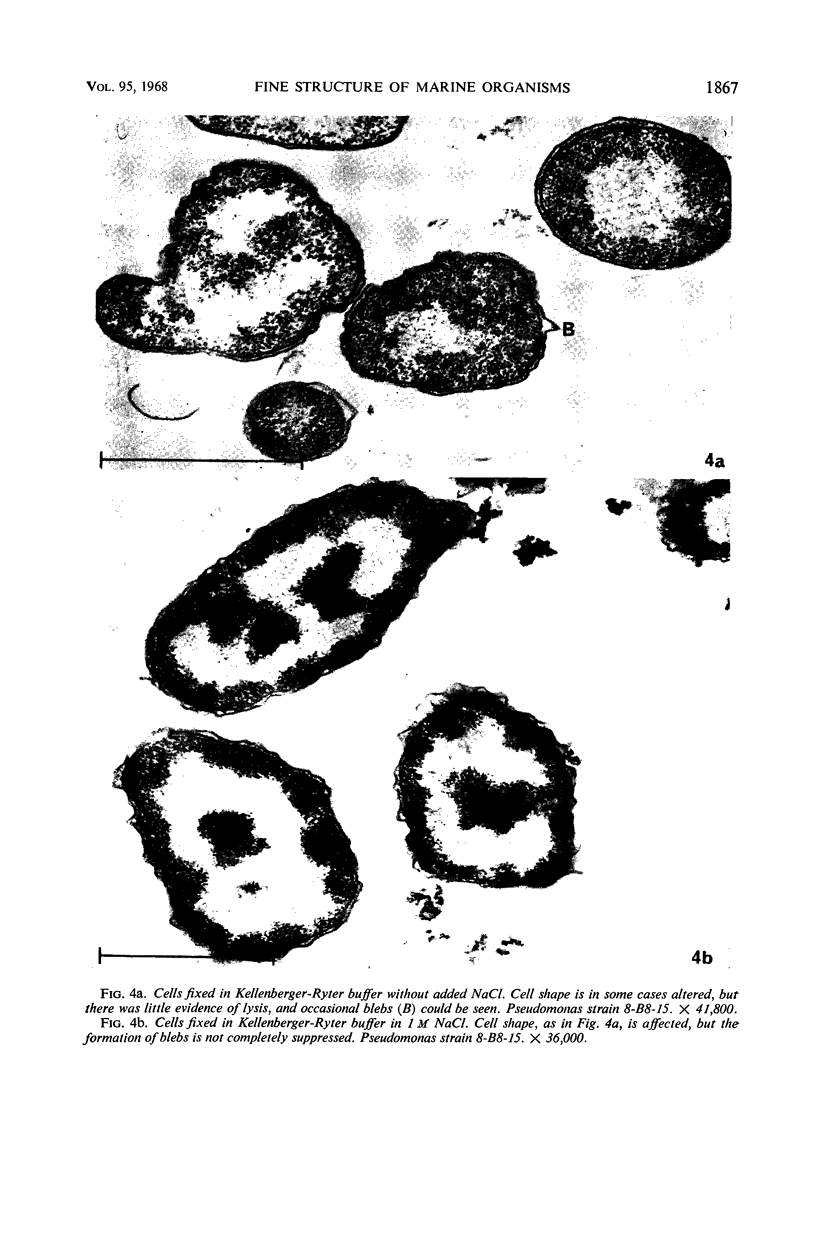
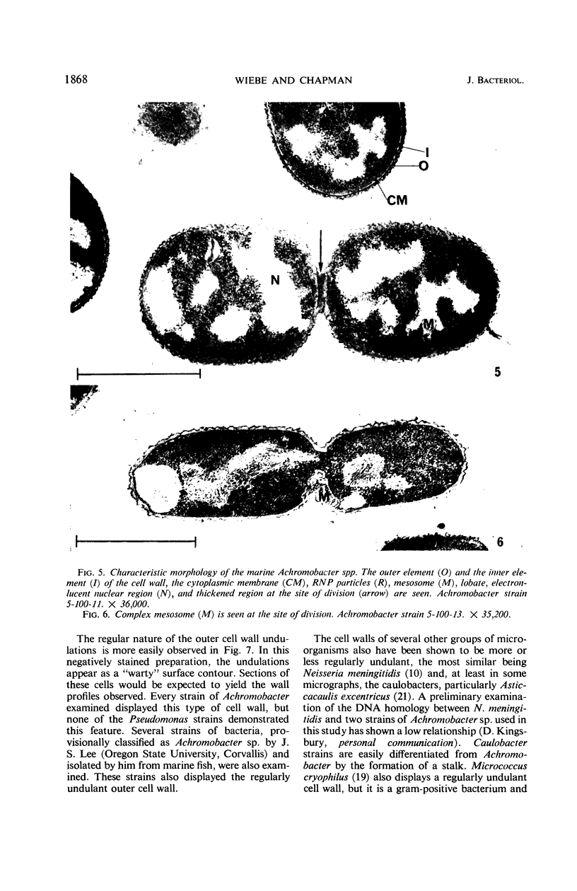
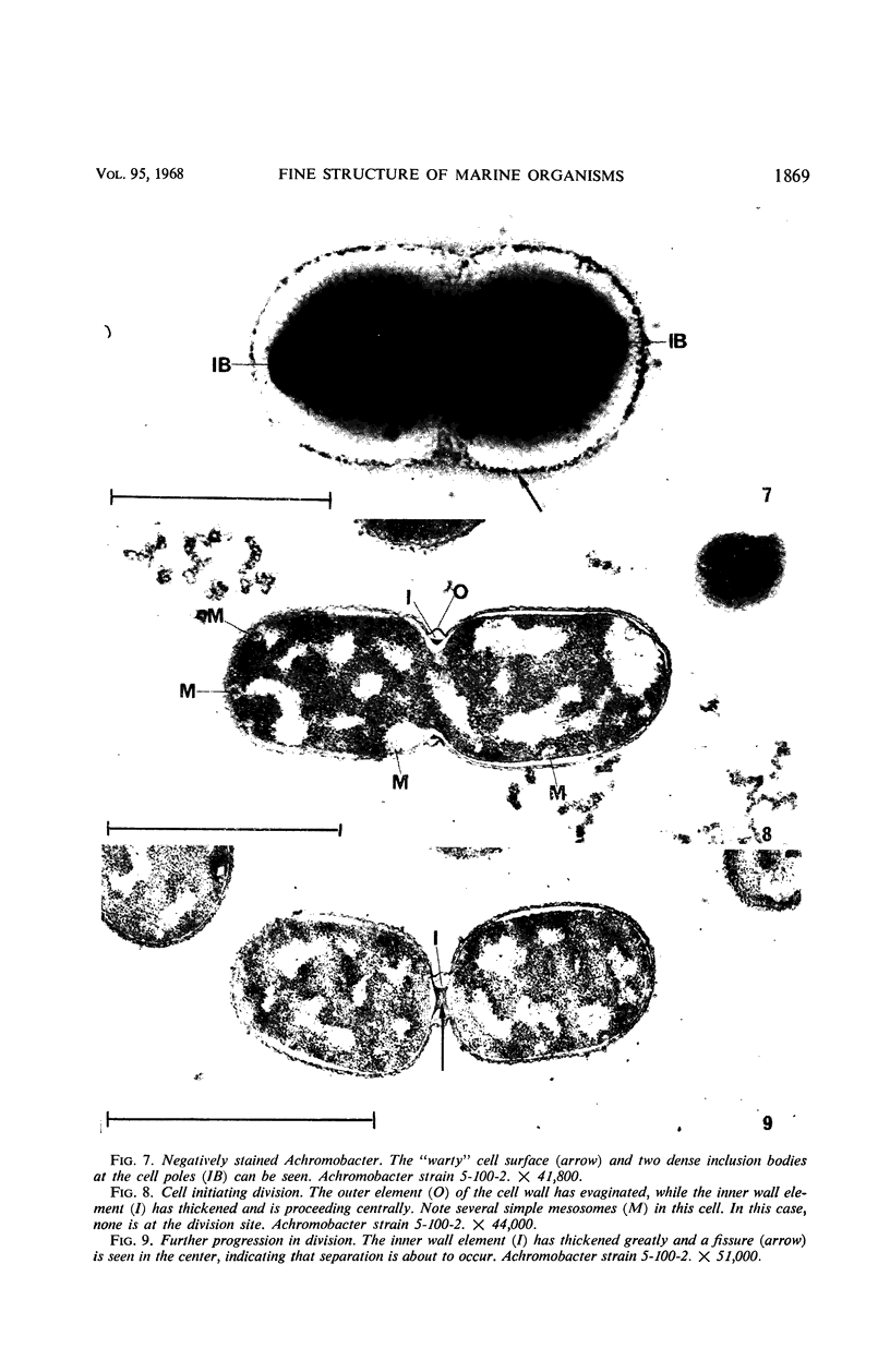
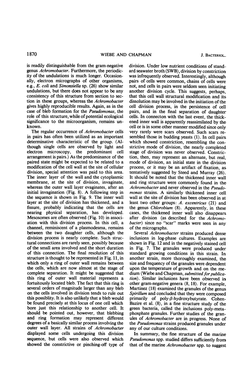
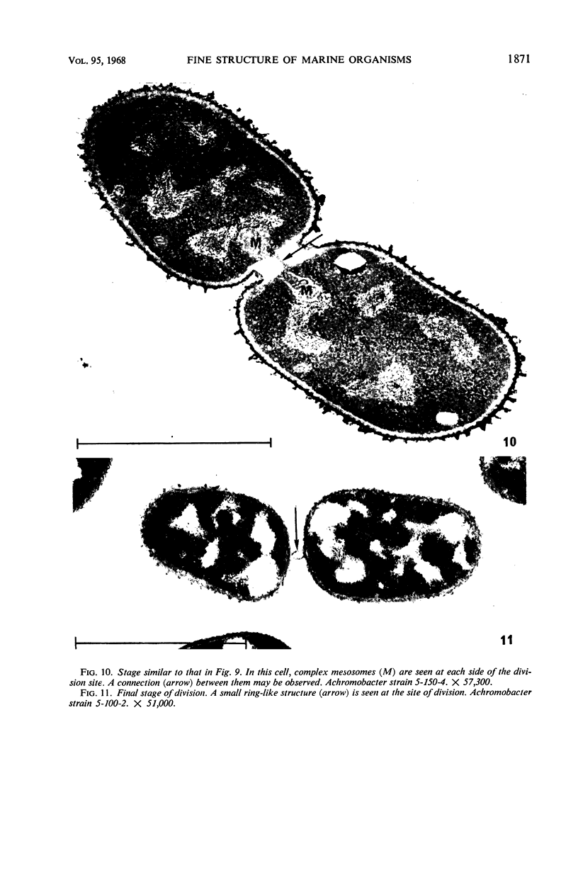
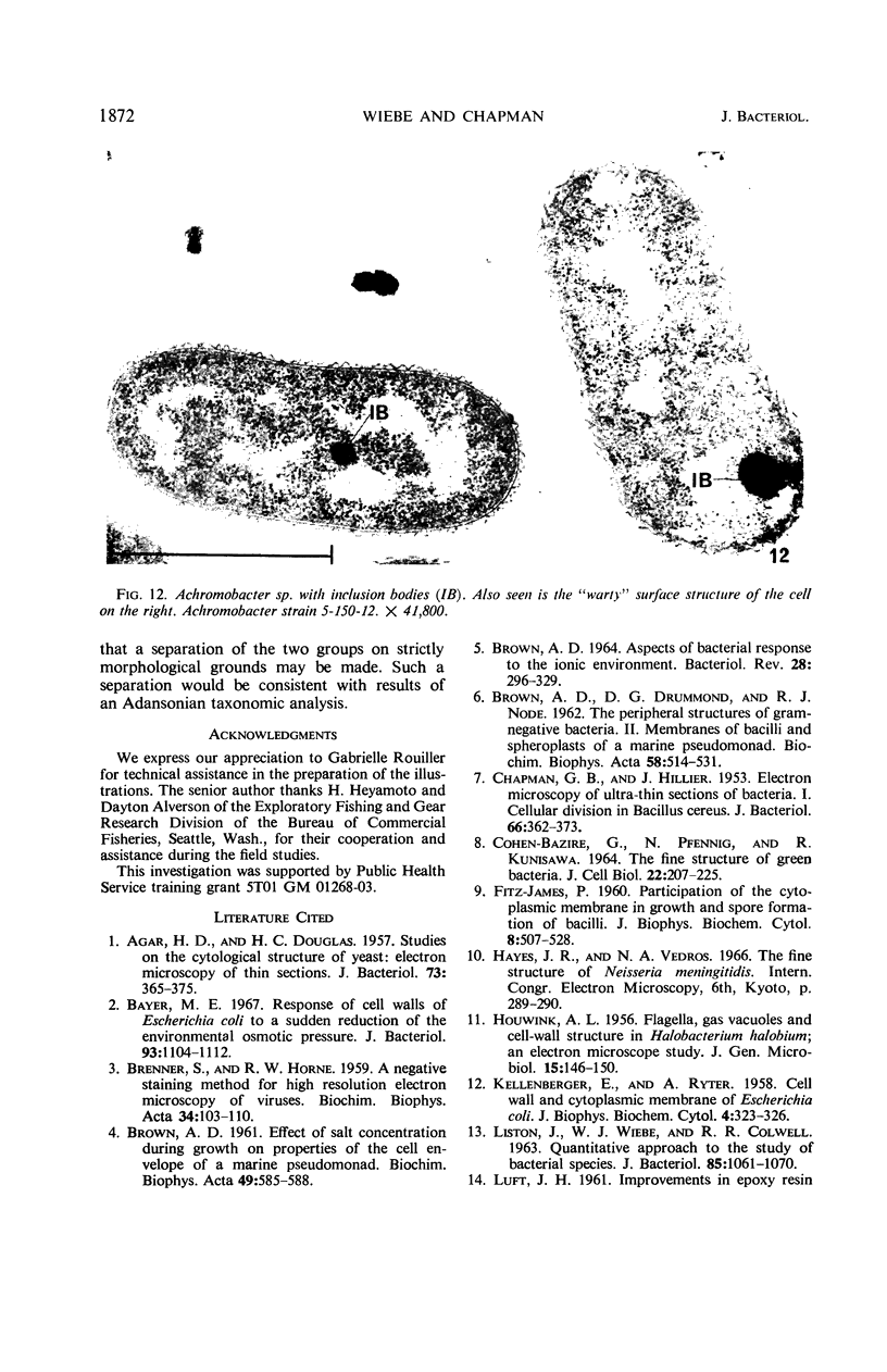
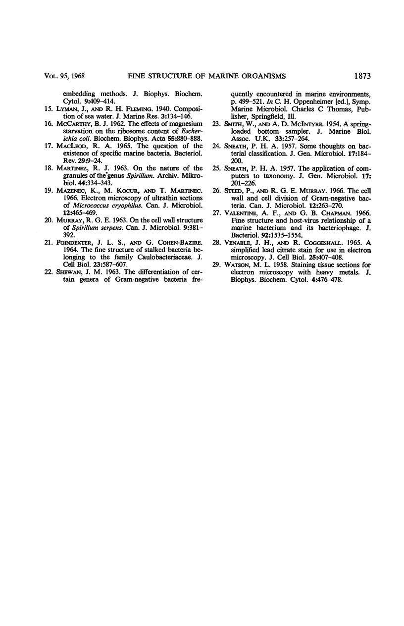
The fine structure of more than 20 marine pseudomonads and more than 15 achromobacters was examined. Under the conditions extant, clear differences between members of these two groups were seen. The pseudomonads displayed the characteristic gram-negative morphology: the cell wall was irregularly undulant and the cytoplasmic membrane more nearly planar, ribonucleoprotein (RNP) particles were loosely packed throughout the periphery of the cytoplasm, and the deoxyribonucleic acid (DNA) was axially disposed. Cell division appeared to be by constriction. Some strains characteristically produced evaginations or blebs of the cell wall. Occasionally, thick, densely stained ring structures were seen which are possibly analogous to mesosomes. In contrast, the achromobacters demonstrated a regularly undulant outer cell wall element and a planar inner wall. The cytoplasmic membrane was thin and not readily observed. RNP particles were densely stained and tightly packed in the cytoplasm; the DNA was most often lobate in disposition. Cellular division was mediated by the formation of a septum which consisted of the cytoplasmic membrane and the inner element of the cell wall. Mesosomes were observed in all of the strains examined. Dense inclusion bodies were also seen in many strains.
Full text
PDF
Images in this article
Selected References
These references are in PubMed. This may not be the complete list of references from this article.
- AGAR H. D., DOUGLAS H. C. Studies on the cytological structure of yeast: electron microscopy of thin sections. J Bacteriol. 1957 Mar;73(3):365–375. doi: 10.1128/jb.73.3.365-375.1957. [DOI] [PMC free article] [PubMed] [Google Scholar]
- BRENNER S., HORNE R. W. A negative staining method for high resolution electron microscopy of viruses. Biochim Biophys Acta. 1959 Jul;34:103–110. doi: 10.1016/0006-3002(59)90237-9. [DOI] [PubMed] [Google Scholar]
- BROWN A. D. ASPECTS OF BACTERIAL RESPONSE TO THE IONIC ENVIRONMENT. Bacteriol Rev. 1964 Sep;28:296–329. doi: 10.1128/br.28.3.296-329.1964. [DOI] [PMC free article] [PubMed] [Google Scholar]
- Bayer M. E. Response of Cell Walls of Escherichia coli to a Sudden Reduction of the Environmental Osmotic Pressure. J Bacteriol. 1967 Mar;93(3):1104–1112. doi: 10.1128/jb.93.3.1104-1112.1967. [DOI] [PMC free article] [PubMed] [Google Scholar]
- CHAPMAN G. B., HILLIER J. Electron microscopy of ultra-thin sections of bacteria I. Cellular division in Bacillus cereus. J Bacteriol. 1953 Sep;66(3):362–373. doi: 10.1128/jb.66.3.362-373.1953. [DOI] [PMC free article] [PubMed] [Google Scholar]
- COHEN-BAZIRE G., PFENNIG N., KUNISAWA R. THE FINE STRUCTURE OF GREEN BACTERIA. J Cell Biol. 1964 Jul;22:207–225. doi: 10.1083/jcb.22.1.207. [DOI] [PMC free article] [PubMed] [Google Scholar]
- FITZ-JAMES P. C. Participation of the cytoplasmic membrane in the growth and spore fromation of bacilli. J Biophys Biochem Cytol. 1960 Oct;8:507–528. doi: 10.1083/jcb.8.2.507. [DOI] [PMC free article] [PubMed] [Google Scholar]
- HOUWINK A. L. Flagella, gas vacuoles and cell-wall structure in Halobacterium halobium; an electron microscope study. J Gen Microbiol. 1956 Aug;15(1):146–150. doi: 10.1099/00221287-15-1-146. [DOI] [PubMed] [Google Scholar]
- KELLENBERGER E., RYTER A. Cell wall and cytoplasmic membrane of Escherichia coli. J Biophys Biochem Cytol. 1958 May 25;4(3):323–326. doi: 10.1083/jcb.4.3.323. [DOI] [PMC free article] [PubMed] [Google Scholar]
- LISTON J., WIEBE W., COLWELL R. R. QUANTITATIVE APPROACH TO THE STUDY OF BACTERIAL SPECIES. J Bacteriol. 1963 May;85:1061–1070. doi: 10.1128/jb.85.5.1061-1070.1963. [DOI] [PMC free article] [PubMed] [Google Scholar]
- LUFT J. H. Improvements in epoxy resin embedding methods. J Biophys Biochem Cytol. 1961 Feb;9:409–414. doi: 10.1083/jcb.9.2.409. [DOI] [PMC free article] [PubMed] [Google Scholar]
- MACLEOD R. A. THE QUESTION OF THE EXISTENCE OF SPECIFIC MARINE BACTERIA. Bacteriol Rev. 1965 Mar;29:9–24. [PMC free article] [PubMed] [Google Scholar]
- Mazanec K., Kocur M., Martinec T. Electron microscopy of ultrathin sections of Micrococcus cryophilus. Can J Microbiol. 1966 Jun;12(3):465–469. doi: 10.1139/m66-067. [DOI] [PubMed] [Google Scholar]
- SNEATH P. H. Some thoughts on bacterial classification. J Gen Microbiol. 1957 Aug;17(1):184–200. doi: 10.1099/00221287-17-1-184. [DOI] [PubMed] [Google Scholar]
- SNEATH P. H. The application of computers to taxonomy. J Gen Microbiol. 1957 Aug;17(1):201–226. doi: 10.1099/00221287-17-1-201. [DOI] [PubMed] [Google Scholar]
- STOVEPOINDEXTER J. L., COHEN-BAZIRE G. THE FINE STRUCTURE OF STALKED BACTERIA BELONGING TO THE FAMILY CAULOBACTERACEAE. J Cell Biol. 1964 Dec;23:587–607. doi: 10.1083/jcb.23.3.587. [DOI] [PMC free article] [PubMed] [Google Scholar]
- Steed P., Murray R. G. The cell wall and cell division of gram-negative bacteria. Can J Microbiol. 1966 Apr;12(2):263–270. doi: 10.1139/m66-036. [DOI] [PubMed] [Google Scholar]
- VENABLE J. H., COGGESHALL R. A SIMPLIFIED LEAD CITRATE STAIN FOR USE IN ELECTRON MICROSCOPY. J Cell Biol. 1965 May;25:407–408. doi: 10.1083/jcb.25.2.407. [DOI] [PMC free article] [PubMed] [Google Scholar]
- Valentine A. F., Chapman G. B. Fine structure and host-virus relationship of a marine bacterium and its bacteriophage. J Bacteriol. 1966 Nov;92(5):1535–1554. doi: 10.1128/jb.92.5.1535-1554.1966. [DOI] [PMC free article] [PubMed] [Google Scholar]
- WATSON M. L. Staining of tissue sections for electron microscopy with heavy metals. J Biophys Biochem Cytol. 1958 Jul 25;4(4):475–478. doi: 10.1083/jcb.4.4.475. [DOI] [PMC free article] [PubMed] [Google Scholar]


